Glaucoma and Glaucoma Eye Problems

Learning about Glaucoma is important for everyone, but especially if anyone in your family has glaucoma or you have known risk factors for Glaucoma.
What Is Glaucoma?
Glaucoma is actually not a single disease but is a group of diseases that can steal your sight without any warning or symptoms. Glaucoma is one of the leading causes of blindness for patients between the ages of 18-65 years of age. It is estimated that approximately 3 million people have Glaucoma, yet only half of those actually know that they have it. While most people are familiar with the eye disease Glaucoma, few are aware of why Glaucoma is such a significant threat to sight. In general, most serious eye diseases or eye conditions produce some symptoms that make patients uncomfortable or disturb their vision. Glaucoma begins without any symptoms or obvious loss of vision. People can actually have rather extensive loss of peripheral vision (i.e. side vision) and not know it. In this way glaucoma is quite insidious and, if not detected, diagnosed and treated early in its course, will lead to progressive and permanent vision loss. This is what makes Glaucoma such a dangerous disease.
The fact that most cases of Glaucoma are initially asymptomatic makes regular screening exams very important. High Intraocular Pressure or IOP (increased pressure within the eye) is one of the diagnostic signs that may indicate the presence of Glaucoma. National studies estimate that between 3-6 million people in the United States have higher than normal Intraocular Pressure (IOP), without obvious clinical signs of damage to the optic nerve. Thus it is likely that there are another million people who may have Glaucoma, but have not yet been diagnosed because they do not have access to eye care or even Glaucoma screenings. Just in the United States, there are approximately 100,000 patients who are believed to be legally blind from Glaucoma.
As we described earlier, the most disturbing attributes of Glaucoma are that in most cases it begins slowly and without visual symptoms. This makes it likely to go unnoticed by patients unless they are consistent about having routine eye examinations with Glaucoma testing. It is entirely possible to have a higher than normal Intraocular Pressure (IOP) and vision loss and simply not know it. Fortunately, Glaucoma is treatable, and in many cases, patients retain excellent vision throughout their lives.


How Often Should I Be Checked for Glaucoma?
At Eyecare Medical Group, we recommend that all patients over 50 years of age who have no previous family history of Glaucoma or other general health conditions such as diabetes or high blood pressure, be evaluated for Glaucoma every two years. If there is any family history of Glaucoma at all, or there any other general health problems, we recommend patients be evaluated for Glaucoma every year beginning at age 40. In addition, we now also know that there is considerable risk for siblings of those who have Glaucoma. In the Nottingham Glaucoma Study, it was found that the siblings of Glaucoma patients are 5 times the risk for developing Glaucoma by the age of 70 and therefore should be examined every year.
Glaucoma is a very complex eye disease and not simply an elevated Intraocular Pressure (IOP). Nonetheless, when detected early it can be successfully treated. Glaucoma specialists provide the full scope of technologically advanced diagnostic testing and treatment, as well as taking the time necessary to give each patient the personal education needed to fully understand their condition and get the best possible outcomes for their patients.
Glaucoma Risk Factors
Most patients-especially if they are over 40 or 50 years of age begin to concern themselves with evaluating their risk for various diseases including Glaucoma. Everyone is at risk for Glaucoma, however, there are a number of factors that by themselves can predispose you to develop Glaucoma, and others that when combined can predispose you to increased risk. They may include:
Types of Glaucoma: Open & Closed Angle Glaucoma
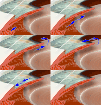
The Glaucoma specialists diagnose and treat a number of types of Glaucoma. To get a better understanding of the different types of Glaucoma, it is necessary to learn about how the normal eye functions. In the “normal” eye, there is a continuous production and drainage of clear colorless fluid called “Aqueous Humor”. This production and drainage is balanced so that an equal amount is produced and drained in order to maintain a “normal’ Intraocular Pressure (IOP).

Aqueous Humor is produced by the Ciliary Body, a structure located just behind Iris, the colored part of the eye that is visible. Normally, the Aqueous Humor is drained through a “filter like” structure called the “Trabecular Meshwork,” which is a tissue meshwork located at the base of the Iris.
In the event that there is either too much Aqueous Humor being produced or too little Aqueous Humor is being drained, there is a rise in pressure inside the eye. This rise in pressure is considered “abnormal.”
There are two main types of Glaucoma: Primary Open Angle Glaucoma (POAG), and Angle Closure Glaucoma. These are characterized by an elevated Intraocular Pressure (IOP), or pressure inside the eye. Sometimes it is possible to have damage to the optic nerve, even with a “normal” Intraocular Pressure. When optic nerve damage has occurred despite a normal IOP, this is called normal-tension Glaucoma.
Secondary Glaucoma refers to any case in which another disease causes or contributes to increased eye pressure, resulting in optic nerve damage and vision loss. As Primary Open Angle Glaucoma and Angle Closure Glaucoma are the most common, we will concentrate on those types of Glaucoma for our discussion.
Primary Open Angle Glaucoma
The most common type of Glaucoma is Primary Open Angle Glaucoma (POAG). Patients with Primary Open Angle Glaucoma usually have an increase in Intraocular Pressure (IOP) upon routine measurement, called Tonometry. This increased Intraocular Pressure (IOP) results from either too much Aqueous Humor being produced or too little being drained as discussed earlier. The fluid buildup within the closed space of the inside of the eye causes the pressure to rise. This elevation in pressure (IOP) can cause permanent damage to the optic nerve resulting in vision loss. The optic nerve is the connection between the retina and the brain and is responsible for communicating visual images. Once the optic nerve is damaged, it is not able to carry visual images, resulting in vision loss. This is why it is so important to monitor, detect and control Intraocular Pressure (IOP). If left untreated, an elevated Intraocular Pressure (IOP) may, over time, cause total blindness.
Angle Closure Glaucoma
Angle Closure Glaucoma can be divided into two main types: Primary Angle Closure Glaucoma and Acute Angle Closure Glaucoma. Although Angle Closure Glaucoma occurs much less frequently than Open Angle Glaucoma, it is important to understand it because it has the ability to produce considerable vision loss in a short period of time. Primary Angle Closure Glaucoma accounts for approximately 10% of all cases of Glaucoma and about 2/3 of these once again produce no early symptoms for patients. Acute Angle Closure Glaucoma is one of the only types of Glaucoma that produce distinct symptoms that include pain, light sensitivity, redness, blurred vision, colored haloes around lights and nausea or vomiting.
Angle Closure Glaucoma is characterized by a blockage or complete closure of the drainage structure of the eye-the Trabecular Meshwork. The Trabecular Meshwork is actually a fine filter. If it is blocked or obstructed by any alteration in the size or shape of the surrounding structures, or by a change in the size or shape of the tissue itself, it will cause the Intraocular Pressure to elevate. In instances where the meshwork becomes blocked abruptly, it will cause a sudden rise in the Intraocular Pressure (IOP), resulting in Acute Angle Closure Glaucoma. Angle Closure Glaucoma is characterized by this sudden rise in pressure which can cause pain, redness, blurred vision and if left untreated permanent loss of vision. While there can be several causes of Angle Closure Glaucoma, it is most often caused by anatomical changes within the internal structures of the eye. Angle Closure Glaucoma is considerably more common in farsighted eyes. Farsighted eyes tend to be smaller. As individuals age, the Crystalline lens gets larger and can obstruct fluid flow, which forces the iris against the trabecular meshwork. The resulting high pressure can damage the optic nerve much like open-angle glaucoma.
During your general eye exam, if your eye doctor observes or measures a narrowed angle, he or she will perform an additional examination procedure called Gonioscopy. This will allow them to fully visualize the meshwork and the angle in order to carefully assess your predisposition to Angle Closure Glaucoma. Gonioscopy is performed by placing a special contact lens on your eye and then using the slit lamp biomicroscope to examine the meshwork and the angle.
In the event that you are at risk for Angle Closure Glaucoma or in the event that you have acute angle-closure glaucoma, your eye doctor may initially prescribe some medication to begin to lower the pressure and then will most likely recommend performing a type of Laser Eye Surgery in order to produce a small opening or hole in the Iris so that Aqueous Humor can quickly and efficiently drain from the eye.
This procedure called “Laser Iridotomy”, is quite successful in reducing the risk of Acute Angle Closure Glaucoma as well as treating Angle Closure Glaucoma. Laser Iridotomy is a type of Laser Eye Surgery that the eye physicians and surgeons and Glaucoma Specialists will perform.
Glaucoma is a very complex eye disease and not simply an elevated Intraocular Pressure (IOP). Nonetheless, when detected early it can be successfully treated. Glaucoma specialists provide the full scope of advanced technology diagnostic testing and treatment, as well as taking the time necessary to give each patient the personal education needed to fully understand their condition and get the best possible outcomes for their patients.
Diagnosis & Tests for Glaucoma
Glaucoma Specialists offer the most advanced diagnostic imaging, diagnostic testing, examination and consultation for the diagnosis of Glaucoma. The key to preventing vision loss from glaucoma is to have regular and complete eye examinations with the necessary Glaucoma diagnostic testing as recommended by an eye doctor. There is a battery of tests that your doctor may use either individually or together in order to make the most precise diagnosis of Glaucoma.
Diagnosis:
If either your Intraocular Pressure (IOP) is elevated or your optic nerve appears unusual, additional tests will be necessary in order to complete the Glaucoma examination. As most types of glaucoma are slowly progressive, it may take years for sufficient evidence to be found, so many of these tests will need to be repeated periodically.
Testing:
During your Glaucoma examination, your eye doctor may perform a Pachymetry Test to measure your corneal thickness as part of your examination and consider this finding in conjunction with the other Glaucoma testing in order to make an accurate diagnosis.
The Pachymetry Test is a simple, quick and painless way of accurately measuring your corneal thickness that our doctors do right in our office. The test is performed by first placing some drops in your eyes to make them numb and then lightly touching the cornea with a small probe that uses sound waves to precisely measure your corneal thickness.
Glaucoma is a very complex eye disease and not simply an elevated Intraocular Pressure (IOP). Nonetheless, when detected early it can be successfully treated. Glaucoma specialists provide the full scope of advanced technology diagnostic testing and treatment, as well as taking the time necessary to give each patient the personal education needed to fully understand their condition and get the best possible outcomes for their patients.


